| 存储条件 |
|---|
| Product Name | CENPB Mouse Monoclonal Antibody |
|---|---|
| Antibody Type | Primary Antibodies |
| Immunogen | Recombinant protein within human CENPB aa 1-200 / 599. |
| Clonality | monoclonal |
|---|---|
| Isotype | IgG2a |
| Host Species | Mouse |
| Tested Applications | ICC/IFIHCWB |
| WB:1:500-1:2000 IHC:1:100-1:500 ICC/IF:1:50-1:100 | |
| Species Reactivity | HumanMouseRat |
| Concentration | 1mg/ml |
| Purification | Protein G |
| Gene Symbol | CENPB |
|---|---|
| Gene Full Name | centromere protein B |
| Gene Summary | This gene product is a highly conserved protein that facilitates centromere formation. It is a DNA-binding protein that is derived from transposases of the pogo DNA transposon family. It contains a helix-loop-helix DNA binding motif at the N-terminus, and a dimerization domain at the C-terminus. The DNA binding domain recognizes and binds a 17-bp sequence (CENP-B box) in the centromeric alpha satellite DNA. This protein is proposed to play an important role in the assembly of specific centromere structures in interphase nuclei and on mitotic chromosomes. It is also considered a major centromere autoantigen recognized by sera from patients with anti-centromere antibodies. [provided by RefSeq, Jul 2008] |
| Molecular Weight(MW) | 65kDa(Observed band size: 77kDa) |
| Cellular Localization | Nucleus, centromere. |
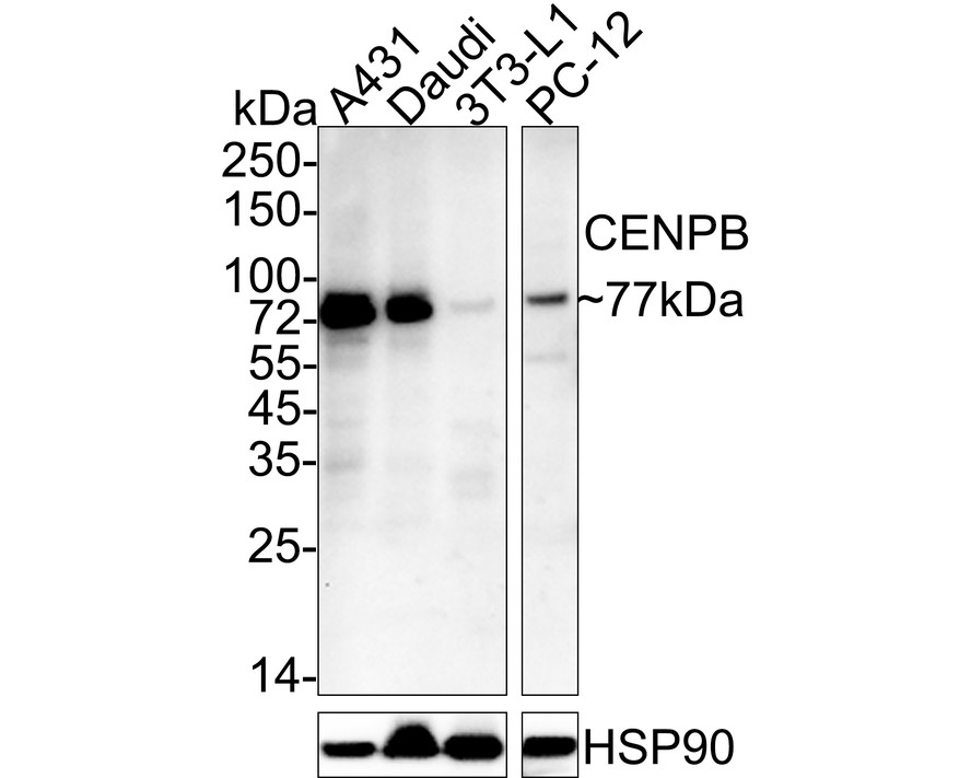
WB
Western blot analysis of CENPB on different lysates with Mouse anti-CENPB antibody at 1/2,000 dilution. Lane 1: A431 cell lysate, Lane 2: Daudi cell lysate, Lane 3: 3T3-L1 cell lysate, Lane 4: PC-12 cell lysate, Lysates/proteins at 30 µg/Lane. Exposure time: 2 minutes; 4-20% SDS-PAGE gel. Proteins were transferred to a PVDF membrane and blocked with 5% NFDM/TBST for 1 hour at room temperature. The primary antibody at 1/2,000 dilution was used in 5% NFDM/TBST at 4℃ overnight. Goat Anti-Mouse IgG - HRP Secondary Antibody at 1/50,000 dilution was used for 1 hour at room temperature.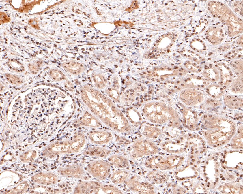
IHC
Immunohistochemical analysis of paraffin-embedded human kidney tissue using anti-CENPB antibody. The section was pre-treated using heat mediated antigen retrieval with sodium citrate buffer (pH 6.0) for 20 minutes. The tissues were blocked in 1% BSA for 30 minutes at room temperature, washed with ddH2O and PBS, and then probed with the primary antibody (1/400) for 30 minutes at room temperature. The detection was performed using an HRP conjugated compact polymer system. DAB was used as the chromogen. Tissues were counterstained with hematoxylin and mounted with DPX.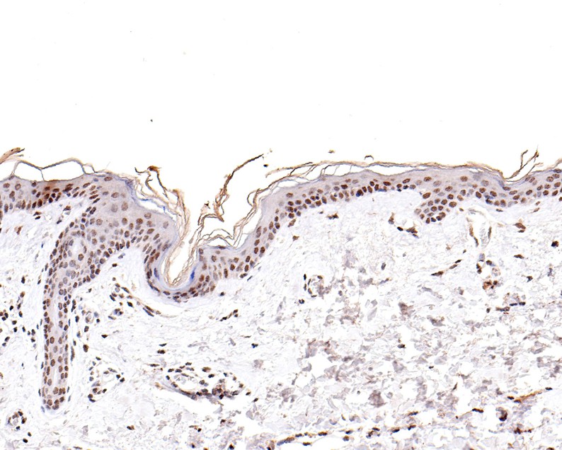
IHC
Immunohistochemical analysis of paraffin-embedded human skin tissue using anti-CENPB antibody. The section was pre-treated using heat mediated antigen retrieval with sodium citrate buffer (pH 6.0) for 20 minutes. The tissues were blocked in 1% BSA for 30 minutes at room temperature, washed with ddH2O and PBS, and then probed with the primary antibody (1/100) for 30 minutes at room temperature. The detection was performed using an HRP conjugated compact polymer system. DAB was used as the chromogen. Tissues were counterstained with hematoxylin and mounted with DPX.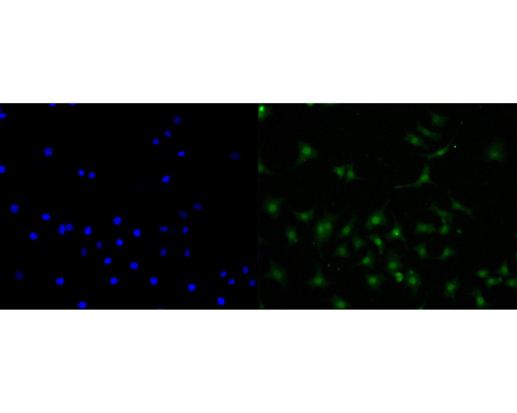
ICC/IF
ICC staining of CENPB in SH-SY5Y cells (green). Methanol fixed cells were blocked with 10% negative goat serum for 15 minutes at room temperature. Cells were probed with the primary antibody (1/50) for 1 hour at room temperature, washed with PBS. Alexa Fluor®488 conjugate-Goat anti-Mouse IgG was used as the secondary antibody at 1/1,000 dilution. The nuclear counter stain is DAPI (blue).| Application Notes | WB:1:500-1:2000 IHC:1:100-1:500 ICC/IF:1:50-1:100 |
|---|
| Form | Liquid |
|---|---|
| Storage Instructions | Store at +4℃ after thawing. Aliquot store at -20℃. Avoid repeated freeze / thaw cycles. |
| Storage Buffer | 1*TBS (pH7.4), 0.2% BSA, 50% Glycerol. Preservative: 0.05% Sodium Azide. |
抱歉,暂无相关文献


