| 存储条件 |
|---|
| Product Name | MAVS Recombinant Rabbit Monoclonal Antibody |
|---|---|
| Antibody Type | Primary Antibodies |
| Immunogen | Synthetic peptide within human MAVS aa 2-50. |
| Clonality | monoclonal |
|---|---|
| Isotype | IgG |
| Host Species | Rabbit |
| Tested Applications | ICC/IFIHCIPWB |
| WB:1:1000 IHC:1:200-1:1000 ICC/IF:1:100 IP:1-2μg/sample | |
| Species Reactivity | HumanMouseRat |
| Concentration | 1mg/ml |
| Purification | Protein A |
| Gene Symbol | MAVS |
|---|---|
| Gene Synonyms | IPS1 VISA IPS-1 CARDIF |
| Gene Full Name | mitochondrial antiviral signaling protein |
| Gene Summary | This gene encodes an intermediary protein necessary in the virus-triggered beta interferon signaling pathways. It is required for activation of transcription factors which regulate expression of beta interferon and contributes to antiviral innate immunity. [provided by RefSeq, Jul 2020] |
| Molecular Weight(MW) | 56kDa(Observed band size: 75kDa) |
| Cellular Localization | Membrane, Mitochondrion, Mitochondrion outer membrane, Peroxisome. |
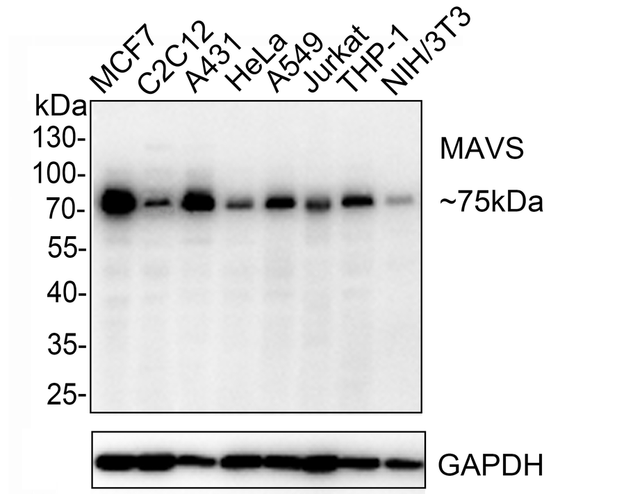
WB
Western blot analysis of MAVS on different lysates with Rabbit anti-MAVS antibody at 1/1,000 dilution. Lane 1: MCF7 cell lysate, Lane 2: C2C12 cell lysate, Lane 3: A431 cell lysate, Lane 4: HeLa cell lysate, Lane 5: A549 cell lysate, Lane 6: Jurkat cell lysate, Lane 7: THP-1 cell lysate, Lane 8: NIH/3T3 cell lysate, Lysates/proteins at 10 µg/Lane. Exposure time: 10S; 10% SDS-PAGE gel. Proteins were transferred to a PVDF membrane and blocked with 5% NFDM/TBST for 1 hour at room temperature. The primary antibody at 1/1,000 dilution was used in 5% NFDM/TBST at room temperature for 2 hours. Goat Anti-Rabbit IgG - HRP Secondary Antibody at 1:200,000 dilution was used for 1 hour at room temperature.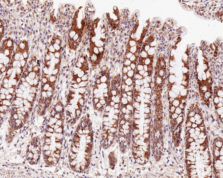
IHC
Immunohistochemical analysis of paraffin-embedded human colon tissue with Rabbit anti-MAVS antibody at 1/200 dilution. The section was pre-treated using heat mediated antigen retrieval with Tris-EDTA buffer (pH 9.0) for 20 minutes. The tissues were blocked in 1% BSA for 20 minutes at room temperature, washed with ddH2O and PBS, and then probed with the primary antibody at 1/200 dilution for 1 hour at room temperature. The detection was performed using an HRP conjugated compact polymer system. DAB was used as the chromogen. Tissues were counterstained with hematoxylin and mounted with DPX.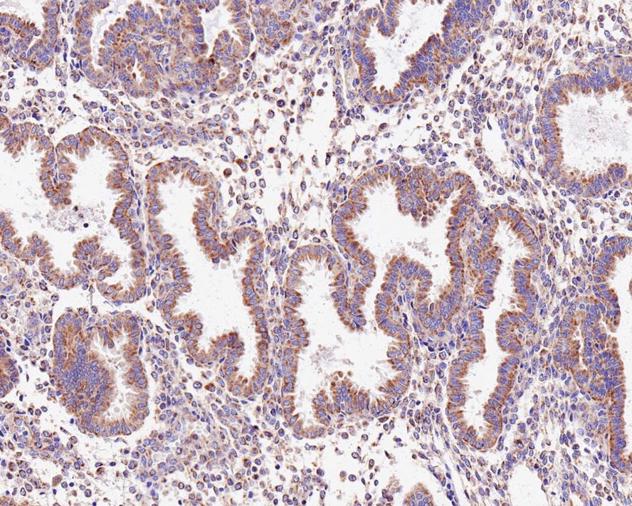
IHC
Immunohistochemical analysis of paraffin-embedded human endometrium tissue with Rabbit anti-MAVS antibody at 1/1,000 dilution. The section was pre-treated using heat mediated antigen retrieval with Tris-EDTA buffer (pH 9.0) for 20 minutes. The tissues were blocked in 1% BSA for 20 minutes at room temperature, washed with ddH2O and PBS, and then probed with the primary antibody at 1/1,000 dilution for 1 hour at room temperature. The detection was performed using an HRP conjugated compact polymer system. DAB was used as the chromogen. Tissues were counterstained with hematoxylin and mounted with DPX.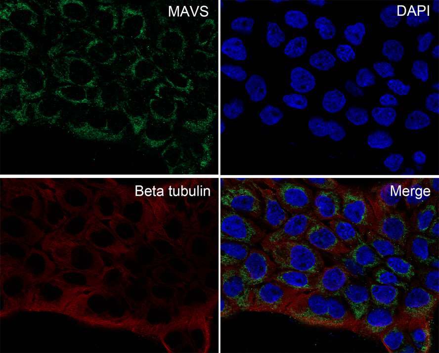
ICC/IF
Immunocytochemistry analysis of A431 cells labeling MAVS with Rabbit anti-MAVS antibody at 1/100 dilution. Cells were fixed in 4% paraformaldehyde for 10 minutes at 37 ℃, permeabilized with 0.05% Triton X-100 in PBS for 20 minutes, and then blocked with 2% negative goat serum for 30 minutes at room temperature. Cells were then incubated with Rabbit anti-MAVS antibody at 1/100 dilution in 2% negative goat serum overnight at 4 ℃. Goat Anti-Rabbit IgG H&L (488) was used as the secondary antibody at 1/1,000 dilution. Nuclear DNA was labelled in blue with DAPI. Beta tubulin (red) was stained at 1/100 dilution overnight at +4℃. Goat Anti-Mouse IgG H&L (647) were used as the secondary antibody at 1/1,000 dilution.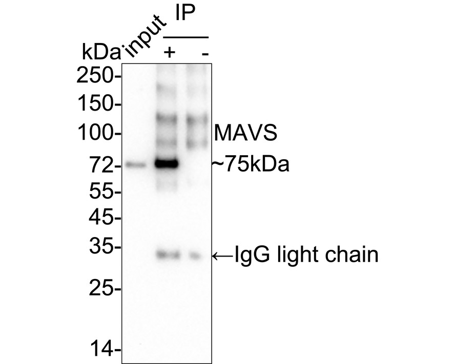
IP
MAVS was immunoprecipitated from 0.2 mg A431 cell lysate with Rabbit anti-MAVS antibody at 2 µg/25 µl agarose. Western blot was performed from the immunoprecipitate using Rabbit anti-MAVS antibody at 1/1,000 dilution. Anti-Rabbit IgG for IP Nano-secondary antibody at 1/5,000 dilution was used for 1 hour at room temperature. Lane 1: A431 cell lysate (input), Lane 2: Rabbit anti-MAVS antibody IP in A431 cell lysate, Lane 3: Rabbit IgG instead of Rabbit anti-MAVS antibody in A431 cell lysate, Blocking/Dilution buffer: 5% NFDM/TBST, Exposure time: 47 seconds.| Application Notes | WB:1:1000 IHC:1:200-1:1000 ICC/IF:1:100 IP:1-2μg/sample |
|---|
| Form | Liquid |
|---|---|
| Storage Instructions | Store at +4℃ after thawing. Aliquot store at -20℃. Avoid repeated freeze / thaw cycles. |
| Storage Buffer | 1*TBS (pH7.4), 0.05% BSA, 40% Glycerol. Preservative: 0.05% Sodium Azide. |
抱歉,暂无相关文献


